I-CAT Cone Beam 3D Dental Imaging System
The current trend in dentistry and dental implantology emphasizes the adoption of three-dimensional X-ray studies as the emerging standard of care. These 3D images furnish crucial diagnostic insights, empowering doctors to plan and execute dental and surgical treatments meticulously.
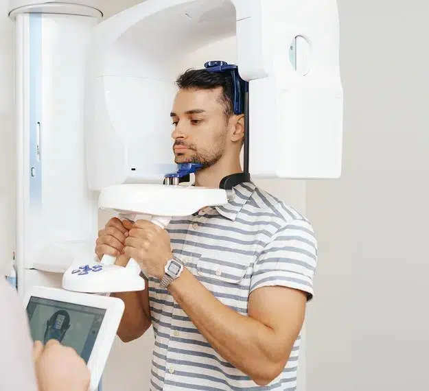
The i-CAT Cone Beam 3D Imaging system brings three-dimensional imaging directly to dental practices. With features like active sagittal, coronal, and axial viewing and manipulation, it significantly enhances diagnosis and treatment planning accuracy. Dental professionals can efficiently utilize the 3D mapping tool to format and select desired slices for immediate examination. Moreover, Cone Beam imaging facilitates rapid and hassle-free image acquisition, with a typical scan completed in just 20 seconds.
The Advantages Of Low-Radiation Systems
The patient benefits from less radiation as well as the comfortable, open environment. Aside from the physical comfort of this system, the doctor can share a visual diagnosis with patients, making them more comfortable with their treatment plan and actively participate in their care. The speed of the scan and the immediate results allows the doctor and patient to better communicate the aspects of a case.
Advantages of i-CAT Over Traditional CT Scans
- Offers advanced imaging within the practice
- Allows doctors to visualize anatomy that can not be diagnosed externally or in 2 dimensional images.
- Safer - delivers less radiation to the patient than traditional CT scans.
- Open environment ensures patient comfort.
- Thorough diagnostic information (optimum view of the critical anatomy of all oral and maxillofacial structures.)
- More accurate treatment planning - confirm certain treatments are necessary.
- More predictable treatment outcomes.
- Reduces the length and cost of therapy.
- Delivers quicker and easier image acquisition - typical scan takes only 20 seconds.
The diagnostic images generated by the i-CAT scanner can seamlessly integrate into third-party software platforms such as Simplant, Nobel Guide, Dolphin, and more. This allows for comprehensive pre-surgical evaluations and facilitates computer-guided surgery. The dentist will have access to a detailed animation, aiding in precise measurement and analysis of your case from all angles. Additionally, the data can be utilized to create customized surgical guides tailored to your specific needs.